

The bead dispenser range
Standard formats from 45 tubes up to 384-well plates, plus fully custom builds.
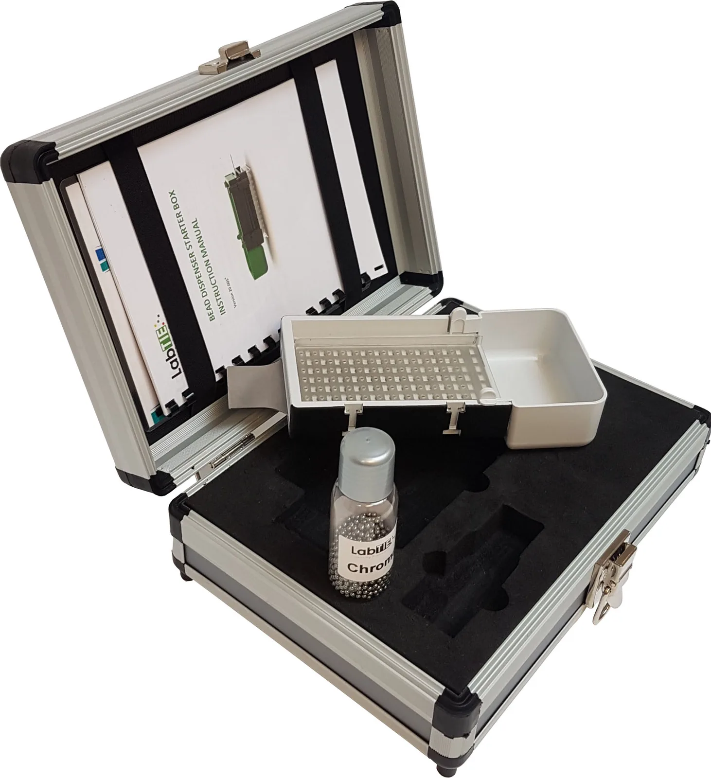
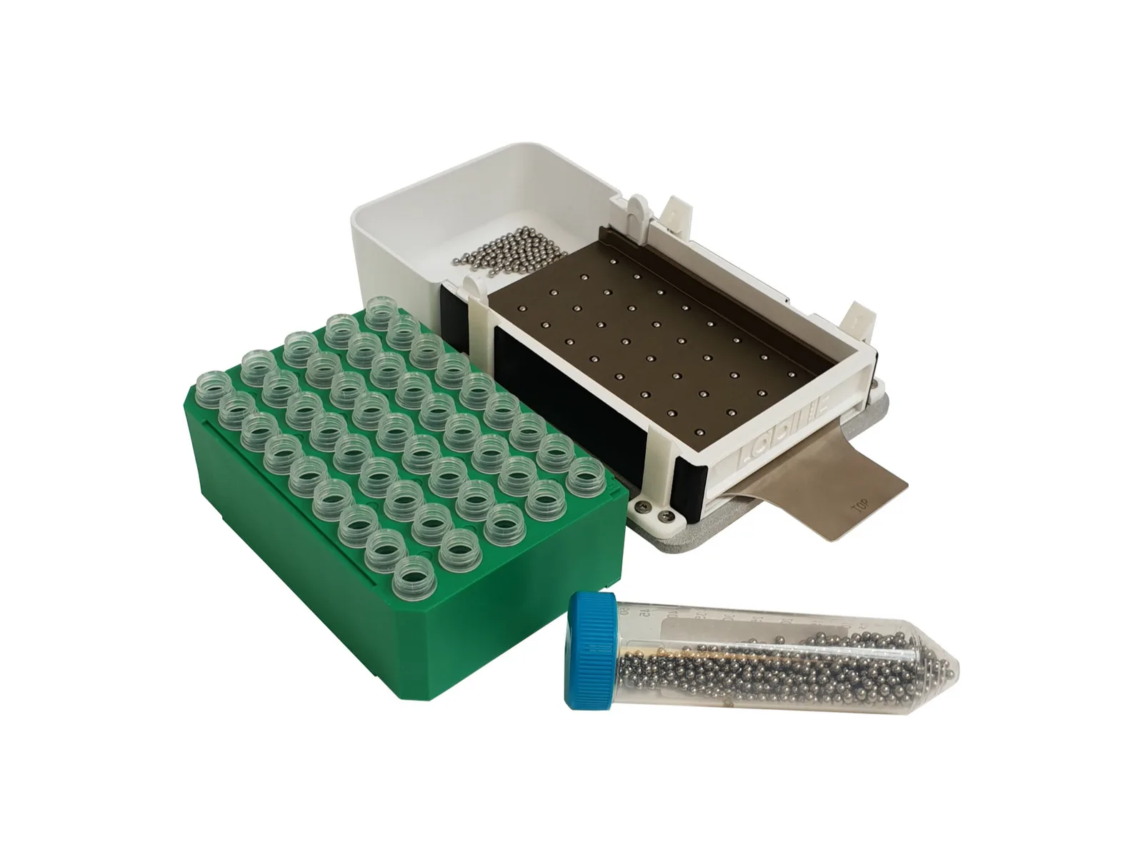


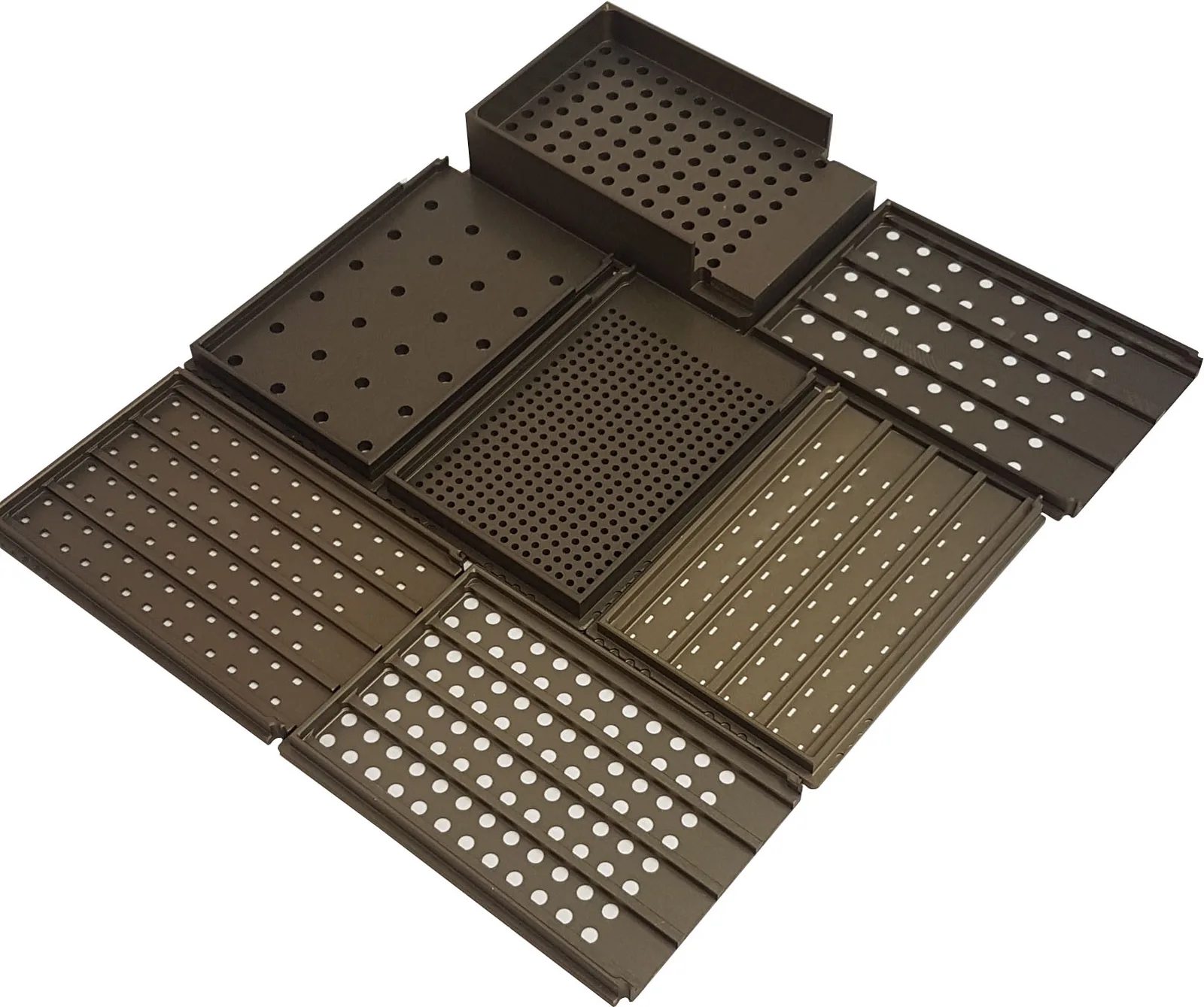
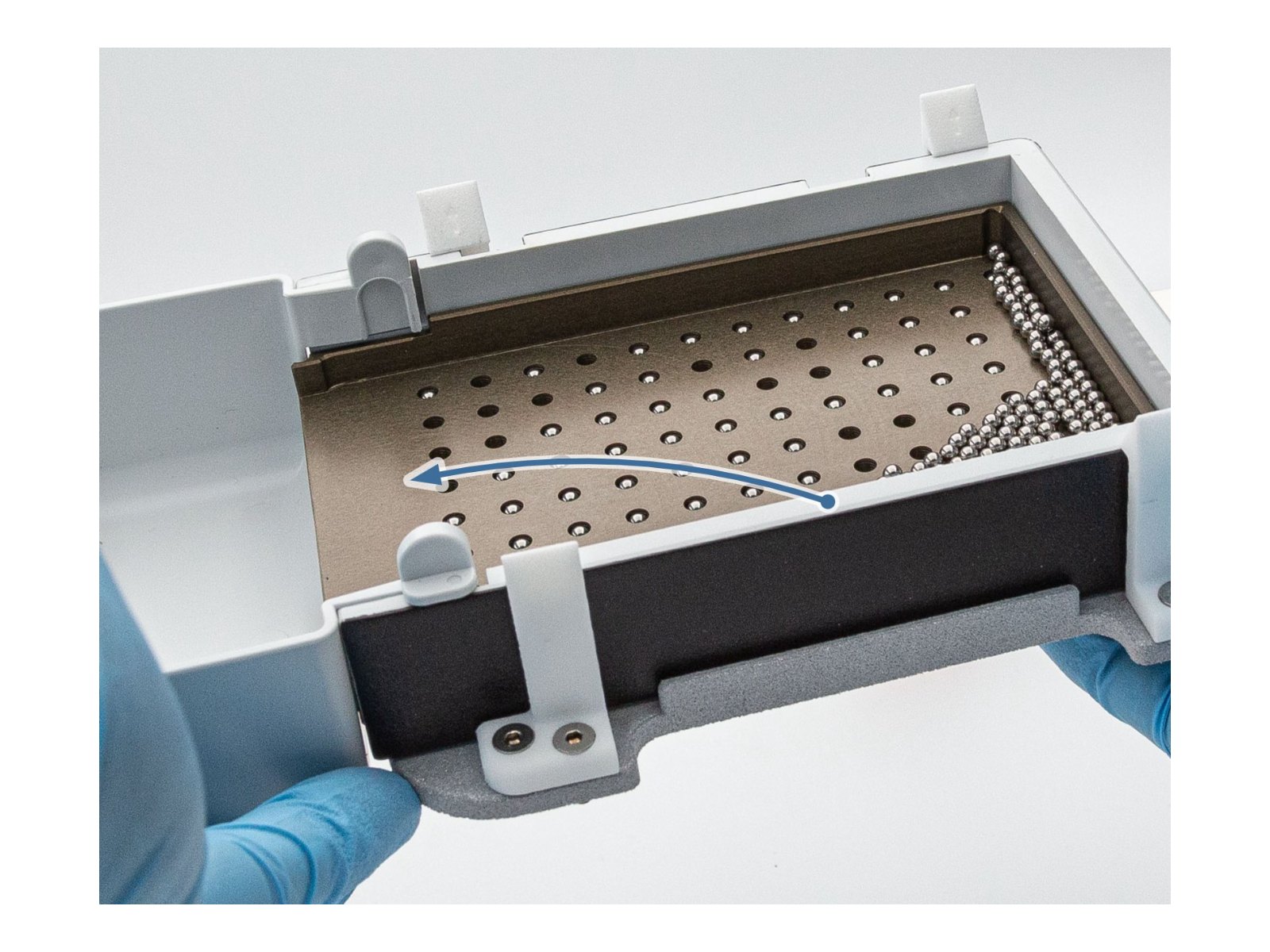
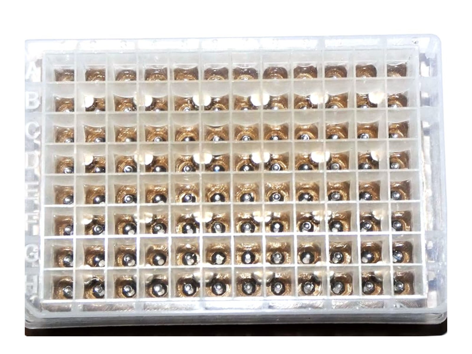
Fill, place, drop
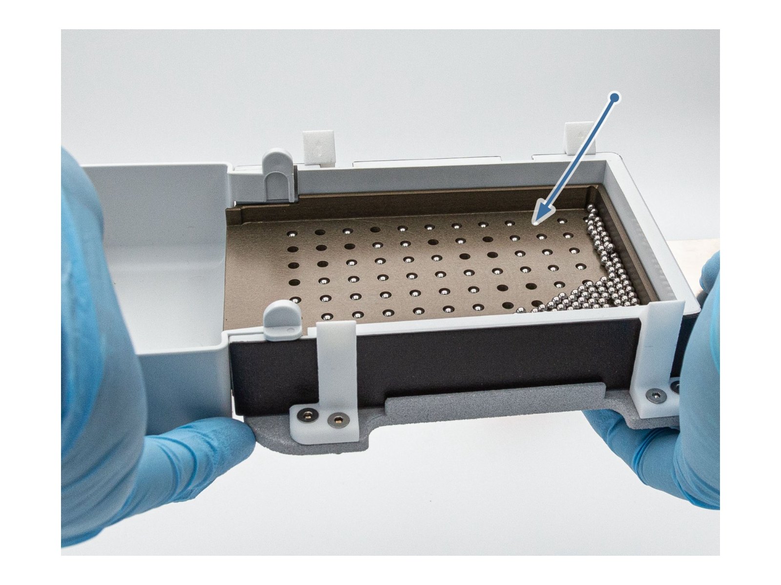

Recommended bead sizes by sample type
Bead size and material must match both the sample and the downstream application. These are practical starting points.

Microbes
Bacteria, yeast and fungi. Cell size sets the scale.
0.2 to 0.5 mm · Yeast: zirconia or silica.
0.5 to 2.8 mm · Moulds and fungi, a size mix usually wins.

Animal tissue
Soft tissue homogenizes with 1 to 3 mm; fibrous tissue needs larger.
Over 100 mg · 5 to 10 mm balls in larger vials.

Plant material
Tough cell walls make plants the most size-sensitive category.
3 to 5 mm · Hard or waxy-cuticle leaves.
3.2 to 7 mm · Seeds and grains.
0.1 to 0.8 mm · Pollen.

Insects
Whole insects carry an exoskeleton and typically need 2 to 4 mm zirconia or steel balls. Under about 5 mg, 2 mm beads lyse well; larger specimens need 5 to 10 mm balls in vials.

Soil and sediment
Heterogeneous, so almost always a bead mix.
0.5 to 1.4 mm · Zirconia for fungi and debris.
2 to 4 mm · Glass or zirconia for aggregates.
Faecal samples
Treat like soil. Tough fibre content calls for a mixed 0.1 to 2.0 mm approach, with zirconia or garnet for extra disruption. Enzymatic pre-treatment is common in microbiome workflows.
Container compatibility: tubes, vials and well plates
Beads must fit and move freely without overfilling the vessel. As a rule, beads should occupy no more than about one third of the volume, since overfilling causes overheating and mechanical failure.
| Vessel | Max bead size | Typical loading |
|---|---|---|
| Microcentrifuge tubes 0.5 to 2.0 ml | 3 mm | 1 to 3 mm beads or a 2.8 mm ball. |
| 24- and 48-well deep plates | About 6 mm | 2 to 4 mm for substantial tissue; 24-well takes 3 to 5 mm balls. |
| 96-well plates | 3 mm | 1 to 2 mm zirconia or a 2.0 to 2.8 mm steel ball. |
| 384-well plates | 2 mm | 0.5 to 1.5 mm zirconia. Below 0.3 mm, use the Powder Dispenser. |
| 4 ml vials | About 8 to 9.5 mm | One 8 mm or 9.5 mm steel ball per 200 mg sample. |
| 15 ml vials | About 11 mm | Two 11 mm steel balls grind roughly 1 g of leaf tissue. |
| Conical tubes 15 / 50 ml | 11 mm and up | Large balls for large volumes, often cryogenic. |
What researchers say
“Ideal for fast filling of steel balls in 96 well format. Filling only a few rows is also possible. The dispenser is easy to use and any surplus of balls is easy to gather again.”
“A tool that must not be missed in any laboratory. It solves the problem of manually dropping and missdropping beads into plates. What used to take minutes is now done in seconds.”
“We searched for a bead dispenser for 96 well plates for over ten years and struggled to find something reliable. What used to be tedious and time consuming is now done in a few seconds.”
Bead beating protocols, bead sizes and materials
Bead beating is a mechanical lysis method that uses small beads, typically 0.1 to 6 mm in diameter, agitated inside tubes or plates to disrupt cells and tissues. Bead selection is not a detail: size, material and hardness must match both the sample and the downstream application.
Read more
Physics of bead impact
When beads are agitated they collide with the sample and transfer kinetic energy on impact. Denser beads carry more energy at the same speed: stainless steel (about 7.9 g/cc) and zirconium oxide (about 6.0 g/cc) impart far more force than equivalent-sized silica glass (about 2.5 g/cc). But small beads move more easily and collide more often, so net disruption depends on both energy per bead and the number of collisions. High-density beads give high-impact collisions but also generate more heat and more nucleic-acid shearing.
Bead material
Low-density beads (glass, silica) are gentle and inert. High-density beads (zirconia, stainless steel, tungsten carbide) are harder and more aggressive. Garnet and silicon carbide are extremely hard and usually combined with zirconia. Zirconia is the right default: a good balance of hardness and inertness. Reach for stainless steel when maximal breakage or total nucleic-acid yield is needed, and avoid it with acids or where metal contamination matters.
DNA, RNA and protein extraction
For genomic DNA, use 1 to 2 mm zirconia or silica plus a couple of 2.8 mm steel balls for tissue, or 0.1 to 0.5 mm silica for bacteria and yeast. Expect fragments in the 2 to 7 kb range. RNA is labile: use 0.5 to 1 mm zirconia or glass, avoid steel or garnet, homogenize quickly in guanidinium buffer and keep tubes cold. For protein, choose medium-hardness zirconia or ceramic at 1 to 3 mm and lyse at 4 °C or in short pulses.
Nucleic acid integrity
Bead beating shears nucleic acids, typically yielding 2 to 7 kb fragments. That is adequate for most PCR and short-read sequencing, and problematic beyond about 10 kb. To preserve length: use larger beads, stop grinding as soon as cells are broken, keep samples cold, and lyse directly into a chaotropic buffer. Where integrity is paramount, grind at cryogenic temperatures instead.
Cleaning and contamination control
Wash beads in warm water with detergent, rinse repeatedly in ultrapure water, dry at 50 to 60 °C until they flow freely, optionally soak in 10% sodium hypochlorite for five minutes to destroy residual DNA and RNA, then autoclave or bake at 180 °C. Beads can usually be reused five to ten times. Discard any that are chipped. For dispensing, prevent static cling of small beads with an anti-static treatment such as nebulized ethanol, and keep mesh plates clean.
Troubleshooting
Low yield: increase bead size or density, add a second size, raise speed or time, and confirm beads occupy about a third of the volume. Homogenate too coarse: move to smaller beads or extend the run. Over-sheared DNA: shorten homogenization, switch to larger or softer beads, pre-chill samples. Heating: use pulse mode, for example 30 s on and 2 min rest, and work on ice. Clogged dispenser: stay above the recommended minimum bead size for the mesh plate, keep sieves clean, and anti-static treat beads before dispensing.
Frequently asked questions
What is a laboratory bead dispenser and how does it work?
A laboratory bead dispenser is a precision tool that loads homogenization or lysis beads into every well, tube or vial in a single motion. LabTIE bead dispensers use an interchangeable mesh plate matched to your bead size: fill the reservoir, tilt so each hole holds exactly one bead, place the dispenser on your plate or rack and pull the slider. A full plate is loaded in 8 to 30 seconds instead of 20 to 30 minutes by hand.
How accurate is a manual bead dispenser?
LabTIE bead dispensers achieve less than 1% placement error across all wells or tubes, rivalling automated liquid-handling systems. The laser-cut mesh plate geometry lets exactly one layer of beads pass per dispensing motion, and consistent bead loading translates directly into more reproducible downstream lysis and extraction results.
Which bead sizes and materials work with LabTIE dispensers?
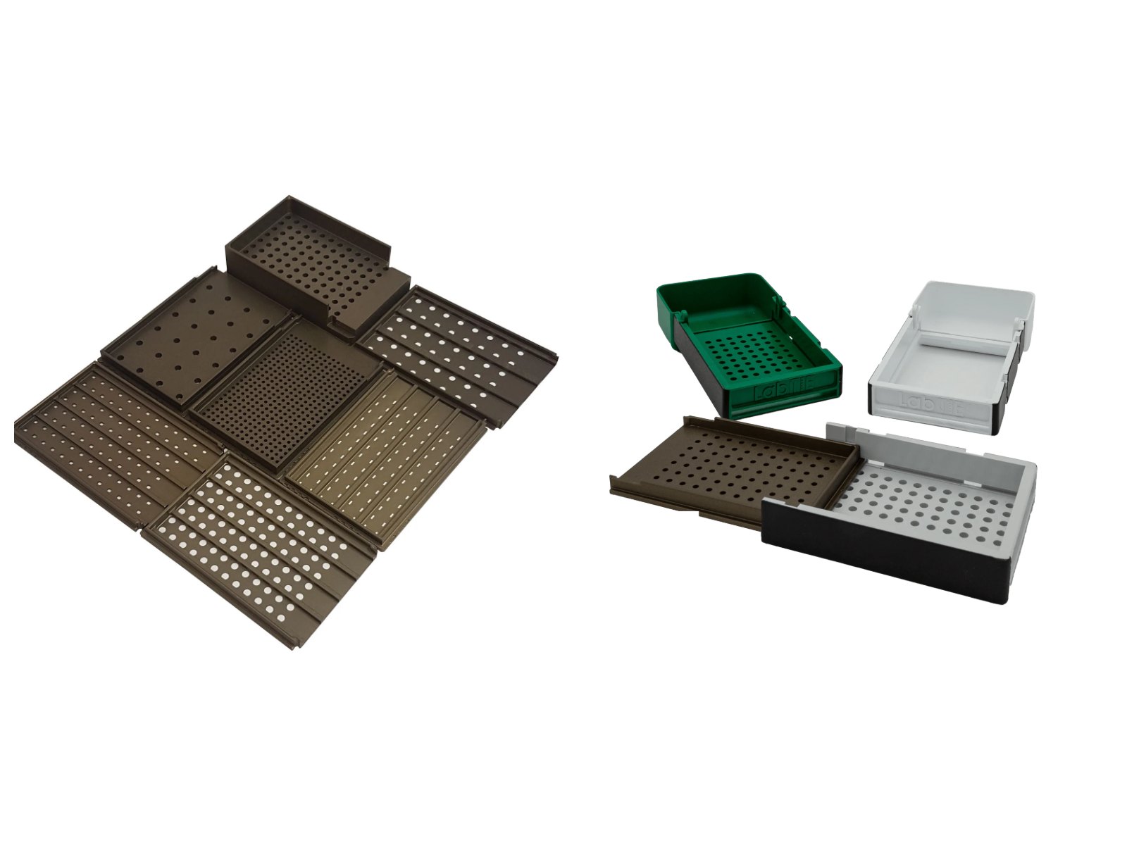
LabTIE dispensers support steel, glass and zirconia beads from 0.3 mm up to 6.0 mm through the interchangeable mesh plate system; common sizes include 0.5, 1.4, 2.0, 2.8, 4.0 and 6.0 mm. Custom mesh plates are available for non-standard bead sizes, and for media finer than 0.3 mm the LabTIE powder dispenser is the right tool.
How do I load beads into a 96-well plate quickly?
With the LabTIE 96-Well Bead Dispenser: fill the reservoir, level the beads so each mesh hole holds one bead, set the dispenser on the plate and pull the slider. All 96 wells are loaded in 8 to 15 seconds with less than 1% error, replacing 20 to 30 minutes of manual bead dropping. The 96-Well Starter kit offers the same core function at a lower entry price.
Can I use a LabTIE bead dispenser for 384-well plates, and how does it differ from the 96-well model?
Yes. The dedicated 384-Well Bead Dispenser loads all 384 positions of an SBS plate in roughly 20 to 30 seconds, using a finer mesh for 0.5 to 1.5 mm zirconia beads. The 96-well model handles larger beads for standard microtiter and deepwell plates. Both share the same interchangeable mesh system and sterilizable design.
How does a bead dispenser improve DNA extraction?
A bead dispenser places a precise number of lysis beads in each well or tube before the bead-beating step, and uniform bead loading is the single most important factor for reproducible DNA yield across a plate. LabTIE dispensers keep well-to-well variability below 1% and support the bead sizes used in commercial extraction kits.
What is the best way to dispense grinding beads for tissue lysis?
Select the LabTIE dispenser that matches your plate or tube format (96-well, 384-well, 48-tube or 45-tube), fit the mesh plate for your bead size, fill the reservoir and pull the slider over your plate or rack. Every position is loaded simultaneously in one smooth motion; more than 120 labs, including at Bayer, Harvard and Syngenta, use this method for consistent bead-based tissue lysis.
How do I choose the right bead dispenser for my lab?
Match the dispenser to your container format first: LabTIE builds models for 96-well plates, 384-well plates, 45-tube racks and 48-tube racks. Then check bead size compatibility via the mesh plates, placement accuracy (target: below 1% error) and sterilizability. The higher your daily sample numbers, the more the 8 to 30 second loading time pays off against manual placement.
How do I clean and sterilize a LabTIE bead dispenser?
All bead-contact components are autoclavable at 121 °C and can be rinsed with 70% ethanol between runs; a simple ethanol wipe is sufficient between sample types. This makes LabTIE dispensers suitable for biological samples under strict contamination-control conditions.
Do LabTIE bead dispensers need electricity or calibration?
No. Every LabTIE bead dispenser is fully mechanical, with no motors, sensors, software or calibration. That makes them practical in biosafety cabinets, GMP-adjacent environments and field labs, with nothing to maintain beyond the exchangeable mesh plate.
Which labs use LabTIE bead dispensers?
More than 120 institutions use LabTIE bead dispensers, including pharmaceutical labs running plate-based lysis in 96-well and 384-well formats, plant genomics and phenotyping groups using plate and tube formats, and organisations such as Harvard, Bayer, Johnson & Johnson, Wageningen University and Syngenta.
Can I order custom mesh plates for non-standard containers?
Yes. LabTIE manufactures custom mesh plates for non-standard plate formats, tube racks and container sizes, and builds fully custom dispensers for proprietary vessels. Send your container dimensions and bead size requirements for a quote and lead time.
What are the shipping times for LabTIE bead dispensers?
LabTIE ships worldwide from the Netherlands. Standard delivery is 3 to 7 business days within Europe and 7 to 14 business days for international orders. Express shipping is available on request at the time of order.
Do you offer a warranty on LabTIE bead dispensers?
Yes. All LabTIE dispensers carry a 12-month warranty covering manufacturing defects under normal laboratory conditions. If a product fails, LabTIE arranges repair or replacement; consumable mesh plates are available separately at low cost.