
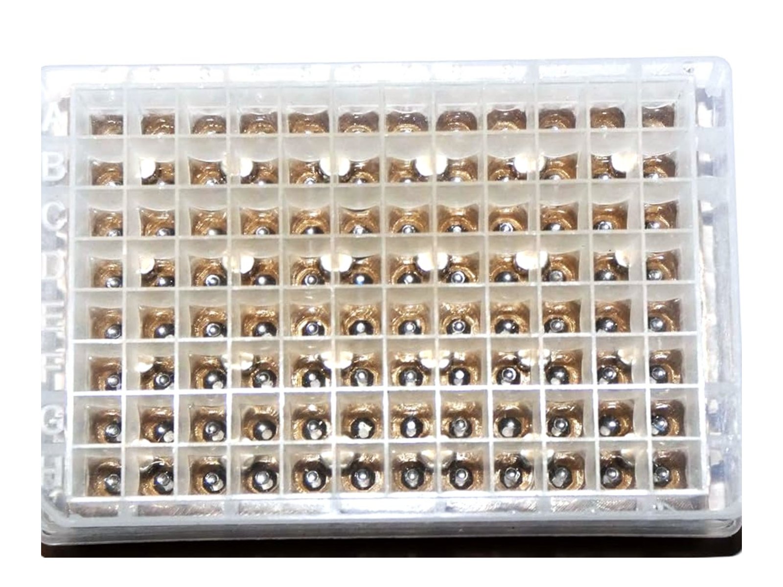
Bead beating usually appears in a methods section as a single line: a bead, an instrument, a number of minutes. Three papers published in 2026 treated that line as something to measure instead of something to copy, and in all three the setting changed the result. Below is what they found about beating time, lysis completeness and instrument choice, and how to check where your own protocol sits.
Too little lysis does not just lower your yield
Mycobacteria have a waxy cell wall that resists chemical lysis. Mulholland and colleagues looked at the ATP burst that mycobacteria appear to produce when treated with cell wall inhibitors, a signal that had been read as a stress response. In the standard whole-cell luminescence assay, which relies on the kit’s own lysis reagent, isoniazid raised the ATP signal 14-fold and 6-fold in two experiments. When the same cultures were opened by bead beating instead, the rise disappeared: ATP was slightly lower in the treated cultures than in the untreated ones, and mass spectrometry agreed. Bead beating untreated cells raised the signal roughly 30-fold over the kit alone, which means the chemical step had been leaving most cells shut (Mulholland CV, Lee BS, Chong A, Cui J, Pethe K, Berney M, 2026, Cell Reports, doi:10.1016/j.celrep.2026.117286).
The drug was not making ATP. It was damaging the wall so the lysis buffer could get in. An incomplete lysis step does not only cost you material: it can produce a number that looks like biology.
Past a certain point, beating cuts the DNA you came for
The opposite error is easier to make, because more beating feels safer. Ali and colleagues were developing host DNA depletion in positive blood cultures for nanopore sequencing, with bead beating on a vortex shaker at 2500 rpm. With the usual 10 minutes of continuous beating, the E. coli sample processed with the salt-activated nuclease was, in their words, highly fragmented, with the longest fragment at 2,110 bp on the TapeStation. Replacing that with three cycles of 2 minutes, each separated by a minute on ice, gave a fragment of 11,604 bp at 0.234 ng/microlitre, and better pathogen read yields downstream (Ali J, Bellankimath AB, Hira J, Chapagain C, Opgard ST, Simonsen GS, Ahmad R, 2026, Frontiers in Microbiology, doi:10.3389/fmicb.2026.1784408).
That is one sample type on one instrument, so the numbers are not a setting you can copy. The direction follows from the mechanism, though: the shear that opens a cell keeps working on the DNA after it is released, and cooling between cycles limits the heat that comes with it. If you need long fragments, beating time is a cost, not a safety margin.
Three minutes on one instrument is not three minutes on another
Jain and colleagues benchmarked five extraction-free lysis devices for tuberculosis detection against a reference method of 95 degrees C for 10 minutes followed by three minutes on a Mini-Beadbeater-16. On sputum swabs in TE buffer, DNA recovery relative to that reference ranged from 11.6 to 53.9 per cent for one device up to 280 to 544 per cent for another; on tongue swabs the spread was narrower but still ran from about 41 to 154 per cent (Jain S, Ball A, Anderson C, Cattamanchi A, Denkinger CM, Steadman A, Yerlikaya S, 2026, medRxiv preprint, doi:10.64898/2026.07.24.26358841).
A preprint has not been through peer review, so treat the individual percentages as provisional. The practical point survives that caveat: a protocol line that reads “bead beat for 3 minutes” does not travel between instruments. Record the instrument, the speed, the cycle structure and the cooling steps, not only the total minutes. Our bead beater protocol guide covers how bead size and speed interact with sample type.
How to find out where your own protocol sits
This takes one plate and one afternoon.
- Take a plate of a sample you run often and give every well the same bead, in the same position.
- Run three conditions, eight wells each: your current time; half that time split into cycles with a minute of cooling between them; and double your current time.
- Measure two things per condition: how much nucleic acid you get, and how long it is, on a gel or a fragment analyser. Add the readout you actually care about, such as Cq or read length.
- Read the pattern. If yield still climbs at the longest setting, you are under-lysing. If yield has flattened while fragment size keeps falling, you are past the useful point.
- Look at the spread across the eight wells, not only the average. Well-to-well spread is what shows up later as an outlier you cannot explain.
That last step only works if the wells started out the same. If one well holds two beads and the next holds none, the spread you measure is loading, not beating.

What published methods hold constant
Two other 2026 papers show how labs pin down the bead itself. A sorghum genotyping study describes the step plainly: tungsten beads, one of 6 mm, were added to each well of a 96-well plate using a bead dispenser after leaf sampling, and the plate was then sealed with silicon caps (Souta CM, Thungo ZG, Sibiya J, Tongooona P, Zikhali M, 2026, Plants, doi:10.3390/plants15162488). A Parkinson’s disease study using a seed amplification assay reports one 3/32 inch silicon nitride bead per well, placed with a house-made bead dispenser, with three technical replicates per sample in 96-well plates (Grillo P, Riboldi GM, Pisani A, Kang UJ, Fereshtehnejad SM, 2026, Journal of Parkinson’s Disease, doi:10.1177/1877718X261417468).
Neither group left the bead to a spatula and a steady hand. One bought a dispenser, the other built one. Either way the bead becomes a fixed part of the method, which is what lets a difference between plates be read as the sample rather than the loading.
That is what our dispensers are for. The 96-Well Bead Dispenser drops beads into all 96 wells in about 8 seconds, with the mesh plate setting the bead size anywhere from 0.8 to 6.0 mm, and the 48 Tube Bead Dispenser does the same for screw cap or snap cap tubes in about 8 seconds. Which bead you load is a separate question; the chrome, stainless steel, zirconia and glass options are listed on the grinding media page, and we wrote about how protocols specify bead number and mass in tissue homogenizer beads: how many per tube or well.
What even loading does not solve
Identical beads in every well do not make lysis complete. A tough sample still needs enough energy and enough cooling, and the Mulholland result is a reminder that a step can look like it worked while most cells are still shut. Uneven sample mass across the plate remains a source of spread, and so does an instrument that does not shake the outer wells as hard as the inner ones. Hand loading adds one more variable on top of those. It is simply the one you can remove first.
The plate machine on our own bench is the Mini-BeadBeater 96, which takes a full deep well plate per run, so the three conditions above can be run side by side on one plate.
Sources
- Mulholland CV, Lee BS, Chong A, Cui J, Pethe K, Berney M (2026). The mycobacterial ATP burst is a lysis artifact and serves as an assay for drug-induced cell wall damage. Cell Reports. doi:10.1016/j.celrep.2026.117286
- Ali J, Bellankimath AB, Hira J, Chapagain C, Opgard ST, Simonsen GS, Ahmad R (2026). Development and optimization of host DNA depletion in blood cultures using a saponin and salt-activated nuclease-based method. Frontiers in Microbiology. doi:10.3389/fmicb.2026.1784408
- Jain S, Ball A, Anderson C, Cattamanchi A, Denkinger CM, Steadman A, Yerlikaya S (2026). Analytical benchmarking of extraction-free lysis devices for molecular detection of tuberculosis. medRxiv preprint. doi:10.64898/2026.07.24.26358841
- Souta CM, Thungo ZG, Sibiya J, Tongooona P, Zikhali M (2026). SNP-based KASP markers reveal genetic diversity and population structure among African sorghum breeding lines and hybrids. Plants. doi:10.3390/plants15162488
- Grillo P, Riboldi GM, Pisani A, Kang UJ, Fereshtehnejad SM (2026). Faster reaction times of CSF alpha-synuclein seed amplification assay predict the diffuse malignant subtype of Parkinson’s disease at 10-year follow-up. Journal of Parkinson’s Disease. doi:10.1177/1877718X261417468
Leave a Reply