
Bead beating is a mechanical cell disruption method that forces small beads against biological samples at high frequency to rupture cell walls and membranes. It is one of the few lysis techniques capable of disrupting gram-positive bacteria, fungal cells, plant tissue, and environmental samples that resist enzymatic or chemical lysis. This guide covers bead size selection by organism, fill volume guidelines, speed and cooling protocols, and troubleshooting for DNA, RNA, and protein extractions.
Choosing the Right Bead Size
Bead size is the single most important parameter in bead beating because it determines collision energy and frequency. For bacteria (0.5 to 5 um cell diameter), 0.1 mm glass or zirconia beads are the standard choice. Their small mass allows thousands of collisions per second, which is the collision frequency that gram-negative organisms require. Switching to 0.5 mm beads reduces that frequency enough to drop lysis efficiency for gram-negatives by 30 to 50%. Gram-positive bacteria, with their thick peptidoglycan walls, benefit from a mixed-bead approach: 0.1 mm beads supply collision frequency while 0.5 mm beads add enough impact energy to crack the wall. Running a 1:1 vol/vol mixture of both sizes consistently outperforms either size alone for species such as Staphylococcus aureus and Mycobacterium.
Larger organisms need proportionally larger beads. Yeast cells (5 to 10 um) lyse efficiently with 0.5 mm glass beads, and switching to zirconia at the same diameter typically raises yield by around 20% because of the higher density and impact force. With optimized settings, 90% lysis is achievable within 5 minutes of total beating time. Tough samples such as plant leaf tissue or woody stem sections require 2.8 mm ceramic or 3.2 mm stainless steel beads. These are heavy enough to fracture cell walls hardened by cellulose and lignin, but should be paired with liquid nitrogen pre-grinding to reduce the sample to a powder before loading the tube. Environmental and soil samples present a mixed community of organism sizes, so combining 0.1 mm and 2.8 mm beads in the same tube covers both the bacterial and larger eukaryotic fractions simultaneously.
| Sample type | Recommended bead | Typical lysis time | Notes |
|---|---|---|---|
| Gram-negative bacteria | 0.1 mm glass or zirconia | 3 to 4 cycles x 45 s | High collision frequency needed |
| Gram-positive bacteria | Mix 0.1 mm + 0.5 mm | 3 to 5 cycles x 60 s | Thick peptidoglycan wall |
| Yeast | 0.5 mm glass (zirconia +20%) | 3 to 4 cycles x 60 s | 90% lysis achievable |
| Plant tissue | 2.8 mm ceramic | 3 cycles x 90 to 120 s | Pre-grind in LN2 recommended |
| Soil / environmental | 0.1 mm + 2.8 mm mix | 4 to 5 cycles x 60 s | Broad cell size coverage |
| Mammalian tissue | 1.4 mm ceramic or steel | 2 to 3 cycles x 30 s | Gentle, to avoid protein denaturation |
Bead Loading and Fill Volume
Fill volume is the second most frequently misoptimized parameter after bead size. The standard guideline is to fill the tube or well 1/3 to 1/2 by volume with beads, then bring the total liquid volume to 70 to 80% of tube capacity with lysis buffer. Underfilling with beads reduces the number of collision events per cycle; overfilling with beads leaves too little headspace for the beads to move freely, which dampens agitation and counterintuitively reduces lysis efficiency. Overfilling with buffer dilutes the lysate and can cause foaming, which both lowers yield and traps nucleic acids at the air-liquid interface where shear forces degrade them faster.
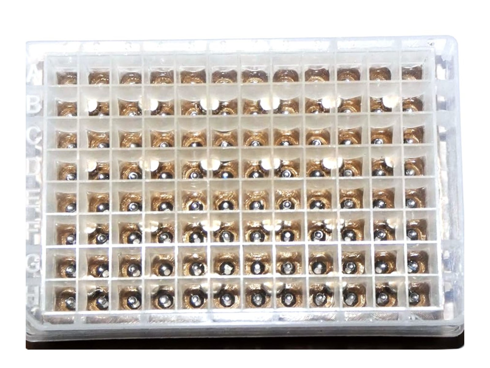
At tube scale, adjusting fill volumes is straightforward. At 96-well plate scale, loading beads manually becomes a significant bottleneck: dispensing beads into 96 individual wells by hand or with a spatula takes 20 to 30 minutes per plate, with well-to-well variation in bead count that directly translates to variation in lysis efficiency across samples. LabTIE bead dispensers solve this by loading all 96 wells in a single slider motion, delivering a controlled, consistent bead count to every well simultaneously and eliminating the manual step entirely.

Speed, Duration, and the Cooling Protocol
Most bead mill homogenizers used in molecular biology operate in the 20 to 30 Hz range. Higher frequency increases kinetic energy per collision, which improves lysis but also generates substantially more heat. Sample temperature rises 5 to 10 degrees Celsius per 60-second cycle at 30 Hz without active cooling. That is consequential: RNA degrades rapidly above 37 degrees Celsius due to RNase activation, and genomic DNA begins to nick mechanically above 60 degrees Celsius. Even for downstream applications that tolerate some DNA fragmentation, heat-induced protein denaturation can co-precipitate nucleic acids and reduce yield. The practical solution is to cap each beating cycle at 45 to 60 seconds, then rest the sample on ice for at least 2 minutes before the next cycle to let the tube return to near 0 degrees Celsius.
The standard bead beating protocol for most molecular biology applications is 30 Hz, 45 to 60 seconds per cycle, 2-minute ice intervals, repeated 3 to 4 cycles. RNA extractions should use 25 Hz with 30 to 45 second cycles and 2 to 3 minutes on ice, because RNA is more sensitive to both heat and mechanical shear than DNA. One underappreciated variable is instrument frequency drift: bead mills that operate continuously over months or years can run 2 to 5 Hz below their set point due to motor wear. If lysis efficiency drops unexpectedly, verify the actual oscillation frequency with a calibration check before troubleshooting beads or buffers.
| Target | Speed | Cycle time | Cycles | Ice interval |
|---|---|---|---|---|
| Genomic DNA | 25 to 30 Hz | 45 to 60 s | 3 to 4 | 2 min on ice |
| Total RNA | 25 Hz | 30 to 45 s | 3 to 5 | 2 to 3 min on ice |
| Total protein | 20 to 25 Hz | 30 s | 4 to 6 | 3 min on ice |
| Metabolomics | 30 Hz | 60 s | 2 to 3 | Dry ice or LN2 |
High-Throughput Bead Beating: 96-Well Scale
Scaling bead beating to 96-well format for high-throughput workflows exposes a bottleneck that is easy to overlook: the instrument run time is not the limiting step. Modern 96-well bead mills such as the TissueLyser II, FastPrep-96, and Precellys Evolution can process a full plate in under 5 minutes. The actual throughput constraint is pre-run plate preparation. Loading beads into 96 wells by hand (with a spatula, a scoop, or a manual pipette) typically takes 20 to 30 minutes per plate, and the well-to-well bead count variation introduced by manual loading creates measurable differences in lysis efficiency across the plate. This variation becomes a significant confounder in microbiome studies, metagenomics workflows, and any application where quantitative consistency between samples matters.
LabTIE bead dispensers address this directly: a single pull of the slider loads all 96 wells with a consistent bead count in one motion, reducing plate prep from 20 to 30 minutes to under a minute and eliminating the inter-well variation that manual loading introduces. The dispensers are compatible with standard deep-well plates used in TissueLyser, FastPrep, Precellys, and other 96-well bead mill formats. For labs running multiple plates per day, the time saving compounds quickly. More information on compatible workflows is available on the sample homogenization page.
Troubleshooting Poor Lysis Efficiency
The most common causes of low lysis yield are wrong bead size for the organism, incorrect fill volume, buffer foaming, and undetected instrument frequency drift. Start by confirming that the bead diameter matches the cell size of your sample: using 0.5 mm beads for gram-negative bacteria or 0.1 mm beads for plant tissue are both common mismatches that produce poor lysis despite correct speed and timing. Check that the tube or well is filled to 1/3 to 1/2 by bead volume, not by weight, and that the total liquid volume does not exceed 80% of tube capacity. Buffer foaming is a sign of over-beating or detergent incompatibility; switching to a lower-surfactant lysis buffer and reducing cycle time by 15 seconds usually resolves it.
For poor DNA quality specifically (low A260/A280, low A260/A230, or smeared gel bands), the most frequent cause is excessive heat from too-long cycles or insufficient ice intervals. Reduce each cycle by 15 seconds and extend ice intervals to 3 minutes. For RNA, RNase contamination is the primary concern: use RNase-free water, tips, and tubes throughout, and reduce beating time to the minimum needed for lysis (30 to 45 s at 25 Hz). If lysis efficiency has dropped gradually over weeks, check instrument calibration before changing the protocol. A 3 to 5 Hz drop in oscillation frequency due to motor wear explains many unexplained yield drops in high-use labs.
Pre-fill bead plates with LabTIE dispensers
Works with TissueLyser, FastPrep, Precellys, and any 96-well bead mill. One slider motion, all wells loaded.
View bead dispensersRelated
The two vial machines this protocol assumes: the Mini-BeadBeater-16 ULTRA at a fixed 3450 strokes per minute, and the Mini-BeadBeater-24 ULTRA with variable speed from 2400 to 3800 rpm and programmable on and off cycling to hold heat down.

Leave a Reply