
What is cell lysis? It is the step that breaks a cell open so that its DNA, RNA or protein can be reached. Everything after it, from the column to the qPCR, depends on how well that step worked. The methods fall into two groups: chemical and enzymatic lysis, which dissolve or digest the cell wall and membrane, and mechanical lysis, which breaks them with force. Three open access papers from 2016 to 2020 put numbers on what the choice does to DNA yield, shearing and the community you end up measuring.
The two families of cell lysis
Chemical lysis uses a detergent such as SDS and a buffer to dissolve the membrane. Enzymatic lysis adds an enzyme such as lysozyme, which digests the bacterial cell wall. Both are gentle, and both work well on cells with a soft wall, such as a bacterial pellet or cultured animal cells.
Mechanical lysis does the breaking with beads. The sample and the beads go into a tube or a well, the instrument shakes them, and the beads hit the cells. It is the method for material that resists chemistry: spores, fungal cells, plant tissue and soil. Our page on cell disruption methods compares these routes by sample type. This article looks at what published comparisons measured.
Bead beating raised DNA yield in rumen samples
Ma and co-workers extracted DNA from 200 µL of rumen fluid from three goats, in triplicate, with five lysis set-ups: a commercial stool kit and a chemical protocol of their own, each with and without bead beating, plus one run with sand instead of beads. Beating used 200 mg of beads or sand in 1 mL of lysis buffer, at 50 Hz for 3 minutes. Bead beating raised total DNA yield (P = 0.001), and sand gave the same yield as beads (P ≥ 0.19) (Ma et al., 2020).
Two details matter for protocol design. The chemical protocol gave less DNA shearing on gels when combined with bead beating than the kit did. And bead beating did not change bacterial richness, but it lowered the number of protozoal sequence variants (P ≤ 0.08). The authors advise against mechanical lysis for protozoal analysis. The same step that helps one group of organisms can cost you another (Ma et al., 2020).
More force is not always more DNA
Gill and co-workers compared four pretreatments before a kit extraction of vaginal samples: 30 minutes of lysozyme, 16 hours of lysozyme, 60 minutes of lysozyme with mutanolysin and lysostaphin, and 30 minutes of lysozyme followed by bead beating. There was no significant difference between methods, except that lysozyme plus bead beating gave a significantly lower DNA yield than the enzyme cocktail or 30 minutes of lysozyme alone (Gill et al., 2016).
The more useful result is about the profile, not the yield. The method explained almost none of the variation between samples (R² = 0.00086), while the sample of origin explained nearly all of it (R² = 0.99). Samples with similar community structure still clustered together whichever method was used. The authors’ advice is plain: use the same lysis method for every sample in a study (Gill et al., 2016). A lysis method that is a little worse but identical across all wells beats a better one that varies.
Soil needs both chemistry and force
Guerra and co-workers tested lysis combinations on clay rich subsoil, using a lysis buffer of 1 M phosphate with 0.5% SDS on 200 mg of dried soil. The best combination was one minute of bead beating followed by 10 minutes of incubation at 65 °C in that buffer. They validated it on five subsoils against a CTAB method and in an interlaboratory comparison, and describe it as reproducible and cost effective (Guerra et al., 2020).
So the question is rarely chemical or mechanical. The best protocol in this work joins a short bead step to a heated buffer. The abstract gives no yield values, so how large the gain is you can only read in the full paper.
Choosing a lysis method for your samples
From these three papers, a short decision rule follows. Start from the cell wall: soft cells may need only buffer or enzyme, and tough or mixed material needs beads. Decide what you must keep intact: long fragments, or a protozoal fraction, argue for shorter or gentler beating. Then fix the protocol and keep it fixed, since the choice of method moved the community profile far less than the choice of sample did.
If you do use beads in a plate, the bead count per well becomes part of the method. We went through how published protocols state it in an article on how many tissue homogenizer beads go per tube or well.
What this means for loading beads
None of the three papers says how the beads got into the tubes, and that is the gap. Equal lysis across a plate assumes equal beads per well. Loading by spoon or by hand varies from well to well, and the papers above give no measurement of how much that matters. We do not claim a number for it here.
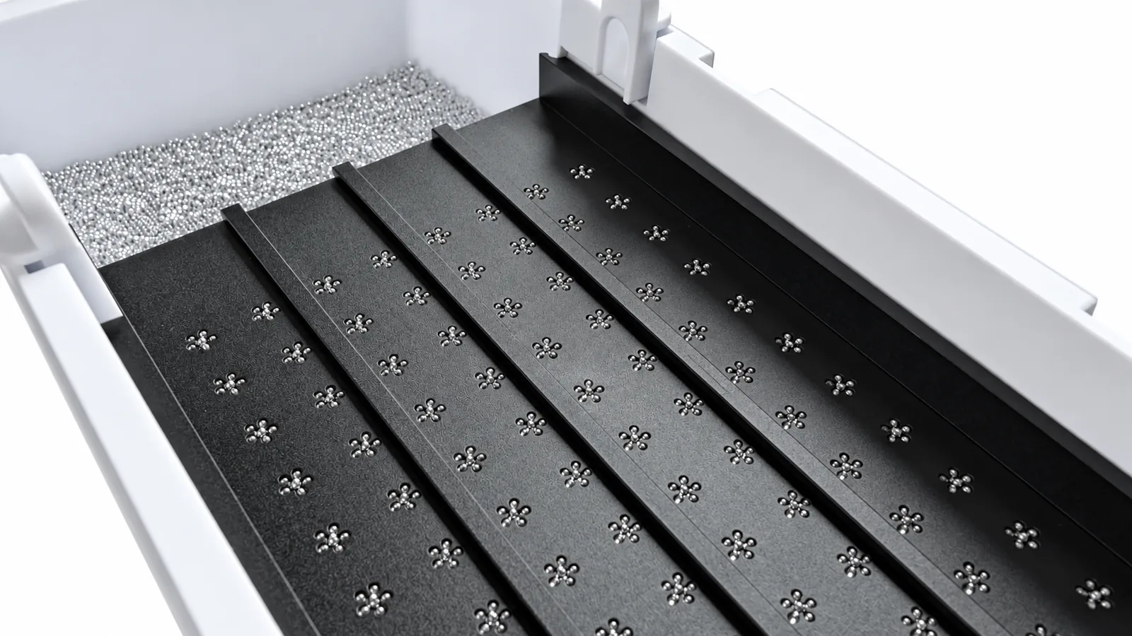
What a dispenser changes is the loading step, not the biology. The 96-Well Bead Dispenser drops beads into all 96 wells of a plate in about 8 seconds, using exchangeable mesh plates, and the bead dispenser range covers tubes and 384 well plates. For the shaking itself, see the bead beaters and the grinding media we supply. A dispenser does not choose your lysis buffer, and it does not fix a protocol that is wrong for your sample.
Limits of this summary
We read the abstracts and the open access article pages, not every table in the supplementary files. The three studies use different samples (rumen fluid, vaginal samples, subsoil), so their numbers do not transfer to your material. They show direction, not a target for your own protocol.
Sources
- Ma ZY, Zhang XM, Wang R, Wang M, Liu T, Tan ZL. Effects of chemical and mechanical lysis on microbial DNA yield, integrity, and downstream amplicon sequencing of rumen bacteria and protozoa. Frontiers in Microbiology. 2020;11. https://doi.org/10.3389/fmicb.2020.581227
- Gill C, van de Wijgert JHHM, Blow F, Darby AC. Evaluation of lysis methods for the extraction of bacterial DNA for analysis of the vaginal microbiota. PLoS ONE. 2016;11(9). https://doi.org/10.1371/journal.pone.0163148
- Guerra V, Beule L, Lehtsaar E, Liao HL, Karlovsky P. Improved protocol for DNA extraction from subsoils using phosphate lysis buffer. Microorganisms. 2020;8(4). https://doi.org/10.3390/microorganisms8040532
Written by Bas Millenaar, LabTIE International B.V., labtie.com. LabTIE builds manual seed, bead and powder dispensers for 96 well plates, 384 well plates, tubes and vials.
Leave a Reply